Contents
How accurate is CT scan for bladder cancer?
Computed tomography (CT) Multidetector (64-slice) CT scanning has provided the mainstay in radiological assessment. It has a reported sensitivity of 85% and specificity of 94% for the diagnosis of bladder cancers [11]. Detection is dependent on the morphology and size of the tumor.
How do they test for bladder cancer?
Cystoscopy. Cystoscopy is the key diagnostic procedure for bladder cancer. It allows the doctor to see inside the body with a thin, lighted, flexible tube called a cystoscope. Flexible cystoscopy is performed in a doctor’s office and does not require anesthesia, which is medication that blocks the awareness of pain.
What can a CT scan of the bladder show?
A CT scan combines x-rays with computer technology to create three-dimensional (3-D) images. These scans can show stones in the urinary tract, as well as obstructions, infections, cysts, tumors, and traumatic injuries. Imaging for urinary stone disease can be done with low or ultra-low dose CT scans.
What is the most reliable test for detecting bladder cancer?
Pathology Tests The most efficient, noninvasive and inexpensive test is a urinalysis/cytology. Here, a sample of urine is taken from the patient and evaluated for cancer cells, red and white blood cells (which fight urinary tract infections), and microscopic hematuria or infection.
What is usually the first symptom of bladder cancer?
In most cases, blood in the urine (called hematuria) is the first sign of bladder cancer. There may be enough blood to change the color of the urine to orange, pink, or, less often, dark red.
What are the warning signs of bladder cancer?
Bladder Cancer: Symptoms and SignsBlood or blood clots in the urine.Pain or burning sensation during urination.Frequent urination.Feeling the need to urinate many times throughout the night.Feeling the need to urinate, but not being able to pass urine.Lower back pain on 1 side of the body.
Can CT scan see inside bladder?
Computed tomography (CT) scan A CT scan uses x-rays to make detailed cross-sectional pictures of your body. A CT scan of the kidney, ureters, and bladder is called a CT urogram. It can provide detailed information about the size, shape, and position of any tumors in the urinary tract, including the bladder.
Why would a urologist order a CT scan?
A CT urogram is used to examine the kidneys, ureters and bladder. It lets your doctor see the size and shape of these structures to determine if they’re working properly and to look for any signs of disease that may affect your urinary system.
Can a CT scan miss a tumor?
Imaging tests usually can’t tell if a change has been caused by cancer. CT scans can produce false negatives and false positives. CT scan can miss cancer, or miss tumors in other areas of the body. CT scans are proven to be less effective at diagnosing cancer than PET/CT.
Is cystoscopy necessary after CT scan?
While some bladder tumors may be found on a CT urogram or other imaging test, others will not. A urologist will often recommend a cystoscopy to evaluate the lower urinary tract (bladder/urethra) for a source of blood in the urine or to workup other urologic symptoms.
Does bladder cancer show up in a blood test?
Tests to diagnose bladder cancer If bladder cancer is suspected, these tests may be performed to diagnose the disease: Physical exam. Blood test: Blood samples are used to measure certain substances released into the blood by organs and tissues in the body.
Will bladder cancer show up in a urine test?
Urinalysis can help find some bladder cancers early, but it has not been shown to be useful as a routine screening test. Urine cytology: In this test, a microscope is used to look for cancer cells in urine. Urine cytology does find some cancers, but it’s not reliable enough to make a good screening test.
Medical History and Physical Exam
Your doctor will want to get your medical history to learn more about your symptoms. The doctor might also ask about possible risk factors, includi…
Transurethral Resection of Bladder Tumor (TURBT)
If an abnormal area (or areas) is seen during a cystoscopy, it will be biopsied to see if it is cancer. A biopsy is the removal of small samples of…
Biopsies to Look For Cancer Spread
If imaging tests suggest the cancer might have spread outside of the bladder, a biopsy might be needed to be sure.In some cases, biopsy samples of…

Imaging Techniques To Detect Bladder Cancer
Imaging techniques, which include ultrasound, computed tomography (or CT) scanning, magnetic resonance imaging (or MRI) and x-ray approaches, provide an important means of assessing the urinary tract, including the kidneys, and play an important role in the detection, diagnosis, and monitoring of bladder cancer.
Detecting bladder cancer with ultrasound
An ultrasound (which may also be referred to as a sonogram) uses high frequency sound waves to produce images of internal organs. Echoes, which are created as sound waves bounce off organs and tissues, produce computer images that provide information on the structure and movement of organs and the blood flow through vessels.
How do ultrasounds help detect and monitor bladder cancer?
An ultrasound of the urinary tract can help assess the size of a bladder tumor and whether a bladder cancer has spread. Ultrasound is able to differentiate between fluid-filled cysts and solid tumors, however, it cannot determine if a tumor is cancerous. Ultrasound can also be used to guide a biopsy needle to sample a suspected cancer.
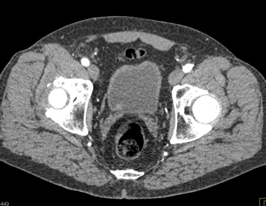
Detecting bladder cancer with CT scans
A CT scan uses x-rays to obtain cross-sectional images of the body. Compared to a general x-ray test, which directs a broad x-ray beam from a single angle, the CT scan uses a number of thin beams to produce a series of images from different angles.
Other imaging approaches to detect or monitor bladder cancer
An MRI scan uses radio waves and magnets to produce more detailed pictures of soft tissues. MRI scans can show whether bladder cancer has spread to other tissues or to the lymph nodes. To improve the quality of the images it’s sometimes necessary to administer an intravenous dye.
What is a CT scan of the bladder?
A CT scan uses x-rays to make detailed cross-sectional pictures of your body. A CT scan of the kidney, ureters, and bladder is called a CT urogram. It can provide detailed information about the size, shape, and position of any tumors in the urinary tract, including the bladder. It can also help show enlarged lymph nodes that might contain cancer, as well as other organs in the abdomen (belly) and pelvis.
How does ultrasound help with bladder cancer?
Ultrasound uses sound waves to create pictures of internal organs. It can be useful in determining the size of a bladder cancer and whether it has spread beyond the bladder to nearby organs or tissues. It can also be used to look at the kidneys. This is usually an easy test to have, and it uses no radiation.
Why is bladder cancer found?
Bladder cancer is often found because of signs or symptoms a person is having. Or it might be found because of lab tests a person gets for another reason. If bladder cancer is suspected, exams and tests will be needed to confirm the diagnosis. If cancer is found, more tests will be done to help find out the extent ( stage) of the cancer.
Can a urine culture show cancer?
If you’re having urinary symptoms, this test may be done to see if an infection (rather than cancer) is the cause. Urinary tract infections and bladder cancers can cause the same symptoms. For a urine culture, a sample of urine is put into a dish in the lab to allow any bacteria that are present to grow. It can take time for the bacteria to grow, so it may take a few days to get the results of this test.
What type of tube is used for bladder cancer?
If bladder cancer is suspected, most doctors will recommend a cystoscopy. . A urologist uses a cystoscope, which is a long, thin, flexible tube with a light and a lens or a small video camera on the end. For details on how this procedure is done, see Cystoscopy.
What is the biopsy for bladder cancer?
A biopsy is when tiny pieces (called samples) of the abnormal-looking tissue are taken out and tested for cancer cells. If bladder cancer is suspected, a biopsy is needed to be sure of the diagnosis.
Is bladder cancer invasive or noninvasive?
This is very important in deciding treatment. If the cancer stays in the inner layer of cells without growing into the deeper layers, it’s called non-invasive. If the cancer grows into the deeper layers of the bladder, it’s called invasive. Invasive cancers are more likely to spread and are harder to treat.
How to diagnose bladder cancer?
This section describes options for diagnosing bladder cancer. Not all tests listed below will be used for every person. Your doctor may consider these factors when choosing a diagnostic test: 1 The type of cancer suspected 2 Your signs and symptoms 3 Your age and general health 4 The results of earlier medical tests
What is the best test for bladder cancer?
There are other urine tests using molecular analysis that can be done to help find cancer, usually at the same time as urinary cytology. Cystoscopy. Cystoscopy is the key diagnostic procedure for bladder cancer. It allows the doctor to see inside the body with a thin, lighted, flexible tube called a cystoscope.
What is the only way to know if you have cancer?
Doctors may also do tests to learn which treatments could work best. For most types of cancer, a biopsy is the only sure way for the doctor to know if an area of the body has cancer. In a biopsy, the doctor takes a small sample of tissue for testing in a laboratory. If a biopsy is not possible, the doctor may suggest other tests …
What is a biopsy of bladder?
A biopsy is the removal of a small amount of tissue for examination under a microscope. This surgical procedure is called a transurethral bladder tumor resection or TURBT. During a TURBT, the doctor removes the tumor and a sample of the bladder muscle near the tumor.
How does radiation work in cancer?
This substance is taken up by cells that use the most energy. Because cancer tends to use energy actively , it absorbs more of the radioactive substance. A scanner then detects this substance to produce images of the inside of the body.
What is a TURBT test?
A TURBT is used to diagnose bladder cancer and find out the type of tumor, how deeply it has grown into the layers of the bladder, and identify any additional microscopic cancerous changes, called carcinoma in situ (CIS) (see Stages and Grades ).
What is PET scan?
However, you may hear your doctor refer to this procedure just as a PET scan. A PET scan is a way to create pictures of organs and tissues inside the body. A small amount of a radioactive substance is injected into the patient’s body.
What is a CT scan?
Computed tomography ( CT) is an imaging procedure that uses special x-ray equipment to create detailed pictures, or scans, of areas inside the body. It is sometimes called computerized tomography or computerized axial tomography (CAT). The term tomography comes from the Greek words tomos (a cut, a slice, or a section) and graphein …
How long does a CT scan last?
The length of a CT procedure depends on the size of the area being scanned, but it usually lasts only a few minutes to half an hour. For most people, the CT is performed on an outpatient basis at a hospital or a radiology center, without an overnight hospital stay.
How old do you have to be to die from lung cancer?
Lung cancer. The NCI-sponsored National Lung Screening Trial (NLST) showed that people aged 55 to 74 years with a history of heavy smoking are 20% less likely to die from lung cancer if they are screened with low-dose helical CT than if they are screened with standard chest x-rays.
What is CAT in medical terms?
It is sometimes called computerized tomography or computerized axial tomography (CAT). The term tomography comes from the Greek words tomos (a cut, a slice, or a section) and graphein (to write or record). Each picture created during a CT procedure shows the organs, bones, and other tissues in a thin “slice” of the body.
How is contrast dye given?
The dye may be given by mouth, injected into a vein, given by enema, or given in all three ways before the procedure. The contrast dye highlights specific areas inside the body, resulting in clearer pictures. Iodine and barium are two dyes commonly used in CT.
Can contrast agents cause kidney problems?
Very rarely, the contrast agents used in CT can cause kidney problems for certain patients, such as those with impaired kidney function. Patient kidney function can be checked with a simple blood test before the contrast agent is injected. CT is a noninvasive procedure and does not cause any pain.
What is the purpose of a biopsy?
To help diagnose the presence of a tumor. To determine exactly where to perform (i.e., guide) a biopsy procedure. To guide certain local treatments, such as cryotherapy, radiofrequency ablation, and the implantation of radioactive seeds. To determine whether a cancer is responding to treatment.