Contents

Can a CT urogram detect bladder cancer?
· Can a CT scan miss bladder cancer? CT scans can provide important information about the urinary tract and bladder tumors. However, while some bladder tumors may be seen on a CT scan, others may not be apparent, such as smaller or flatter tumors. What to expect when having a CT scan. A CT scan is a painless procedure that is typically performed on an …
Can an abdomen CAT scan diagnose bladder cancer?
· So, a doctor’s usage of a CT Scan to detect bladder cancer may show certain things. This may include the following: The formation of stones in the urinary tract can be seen. Infections; Cysts; Obstructions; Tumors, and; Traumatic Injuries; However, a doctor can use a low or high-dose CT Scan to trace urinary stones. Why would a nephrologist ask for CT Scan?
Does cancer always show up in a CT scan?
If an abdominal CT scan shows a normal bladder, don’t celebrate yet. But if it comes back indicating cancer, don’t panic yet, either. About 80,000 people in the U.S. are diagnosed with bladder cancer every year. The five year survival rate, on average, is about 76.8 percent. This not-so-good survival rate is a function of the disease being caught at a later stage than it is of just …
Can a CT scan determine a problem with the gallbladder?
A CT scan of the kidney, ureters, and bladder is called a CT urogram. It can provide detailed information about the size, shape, and position of any tumors in the urinary tract, including the bladder. It can also help show enlarged lymph nodes that might contain cancer, as well as other organs in the abdomen (belly) and pelvis.

Can bladder cancer be missed on CT scan?
Can a CT scan miss bladder cancer? CT scans can provide important information about the urinary tract and bladder tumors. However, while some bladder tumors may be seen on a CT scan, others may not be apparent, such as smaller or flatter tumors.
How accurate is a CT scan for bladder cancer?
Computed tomography (CT) Multidetector (64-slice) CT scanning has provided the mainstay in radiological assessment. It has a reported sensitivity of 85% and specificity of 94% for the diagnosis of bladder cancers [11]. Detection is dependent on the morphology and size of the tumor.
How do you rule out bladder cancer?
Tests for bladder cancer look for different substances and/or cancer cells in the urine. Urinalysis: One way to test for bladder cancer is to check for blood in the urine ( hematuria). This can be done during a urinalysis, which is a simple test to check for blood and other substances in a sample of urine.
What will a CT scan of the bladder show?
A CT scan combines x-rays with computer technology to create three-dimensional (3-D) images. These scans can show stones in the urinary tract, as well as obstructions, infections, cysts, tumors, and traumatic injuries. Imaging for urinary stone disease can be done with low or ultra-low dose CT scans.
Where does it hurt if you have bladder cancer?
Bladder cancer can cause lower back pain when it reaches a more advanced form of the disease. The pain is typically only on one side of the back, but it can be centrally located. Lower back pain might occur once the tumors increase in size or cancer cells start to spread to other parts of your body.
How do I find out if I have bladder cancer?
Tests for bladder cancer look for different substances and/or cancer cells in the urine. Urinalysis: One way to test for bladder cancer is to check for blood in the urine ( hematuria). This can be done during a urinalysis, which is a simple test to check for blood and other substances in a sample of urine.
What is usually the first symptom of bladder cancer?
In most cases, blood in the urine (called hematuria) is the first sign of bladder cancer. There may be enough blood to change the color of the urine to orange, pink, or, less often, dark red.
What are the symptoms of stage 1 bladder cancer?
SymptomsBlood in urine (hematuria), which may cause urine to appear bright red or cola colored, though sometimes the urine appears normal and blood is detected on a lab test.Frequent urination.Painful urination.Back pain.
Does bladder cancer feel like a UTI?
Bladder cancer can be mistaken for a Urinary Tract Infection (UTI) because many of the symptoms overlap. Patients may experience increased frequency and urgency of urination, pain with urination, or urinary incontinence.
Can a CT scan miss a tumor?
Imaging tests usually can’t tell if a change has been caused by cancer. CT scans can produce false negatives and false positives. CT scan can miss cancer, or miss tumors in other areas of the body. CT scans are proven to be less effective at diagnosing cancer than PET/CT.
Can CT scan see inside bladder?
Computed tomography (CT) scan A CT scan uses x-rays to make detailed cross-sectional pictures of your body. A CT scan of the kidney, ureters, and bladder is called a CT urogram. It can provide detailed information about the size, shape, and position of any tumors in the urinary tract, including the bladder.
Why would a urologist order a CT scan?
A CT urogram is used to examine the kidneys, ureters and bladder. It lets your doctor see the size and shape of these structures to determine if they’re working properly and to look for any signs of disease that may affect your urinary system.
Medical History and Physical Exam
Your doctor will want to get your medical history to learn more about your symptoms. The doctor might also ask about possible risk factors, includi…
Transurethral Resection of Bladder Tumor (TURBT)
If an abnormal area (or areas) is seen during a cystoscopy, it will be biopsied to see if it is cancer. A biopsy is the removal of small samples of…
Biopsies to Look For Cancer Spread
If imaging tests suggest the cancer might have spread outside of the bladder, a biopsy might be needed to be sure.In some cases, biopsy samples of…
How many people are diagnosed with bladder cancer every year?
If an abdominal CT scan shows a normal bladder, don’t celebrate yet. But if it comes back indicating cancer, don’t panic yet, either. About 80,000 people in the U.S. are diagnosed with bladder cancer every year. The five year survival rate, on average, is about 76.8 percent.
What is the survival rate for bladder cancer?
The five year survival rate, on average, is about 76.8 percent. This not-so-good survival rate is a function of the disease being caught at a later stage than it is of just a hard-to-treat cancer. About four times more men get bladder cancer than do women.
What is a cystoscope?
It’s a diagnostic procedure (performed either under local anesthetic or general anesthesia) during which a doctor inserts a cystoscope (hollow tube) equipped with a lens into the urethra and further into the bladder.
Can a CT scan detect a UTI?
“CT scan is able to detect large bladder irregularities, but not always small lesions,” says Dana Rice, MD, a board certified urologist and creator of the UTI Tracker mobile app, which helps patients catalog daily urinary tract symptoms, medication and behavioral patterns, and offers personalized tips for UTI prevention.
What is the best way to diagnose bladder cancer?
Cystoscopy. If bladder cancer is suspected, most doctors will recommend a cystoscopy. . A urologist uses a cystoscope, which is a long, thin, flexible tube with a light and a lens or a small video camera on the end. For details on how this procedure is done, see Cystoscopy.
What tests are used to check for bladder cancer?
These include the tests called NMP22 ® (or BladderChek ® ), BTA Stat ®, Immunocyt ® , and UroVysion ®, which are discussed in Can Bladder Cancer Be Found Early?
What is the blue light in a cystoscopy?
Fluorescence cystoscopy (also known as blue light cystoscopy) may be done along with routine cystoscopy. For this exam, a light-activated drug is put into the bladder during cystoscopy. It’s taken up by cancer cells. When the doctor then shines a blue light through the cystoscope, any cells containing the drug will glow (fluoresce). This can help the doctor see abnormal areas that might have been missed by the white light normally used.
What is the biopsy for bladder cancer?
A biopsy is when tiny pieces (called samples) of the abnormal-looking tissue are taken out and tested for cancer cells. If bladder cancer is suspected, a biopsy is needed to be sure of the diagnosis.
What is a physical exam for bladder cancer?
A physical exam can provide information about possible signs of bladder cancer and other health problems. The doctor might do a digital rectal exam (DRE), during which a gloved, lubricated finger is put into your rectum. If you are a woman, the doctor might do a pelvic exam as well.
How long does it take for a urine culture to show up?
It can take time for the bacteria to grow, so it may take a few days to get the results of this test.
How does ultrasound help with bladder cancer?
Ultrasound uses sound waves to create pictures of internal organs. It can be useful in determining the size of a bladder cancer and whether it has spread beyond the bladder to nearby organs or tissues. It can also be used to look at the kidneys. This is usually an easy test to have, and it uses no radiation.
Imaging Techniques To Detect Bladder Cancer
Imaging techniques, which include ultrasound, computed tomography (or CT) scanning, magnetic resonance imaging (or MRI) and x-ray approaches, provide an important means of assessing the urinary tract, including the kidneys, and play an important role in the detection, diagnosis, and monitoring of bladder cancer.
Detecting bladder cancer with ultrasound
An ultrasound (which may also be referred to as a sonogram) uses high frequency sound waves to produce images of internal organs. Echoes, which are created as sound waves bounce off organs and tissues, produce computer images that provide information on the structure and movement of organs and the blood flow through vessels.
How do ultrasounds help detect and monitor bladder cancer?
An ultrasound of the urinary tract can help assess the size of a bladder tumour and whether a bladder cancer has spread. Ultrasound is able to differentiate between fluid-filled cysts and solid tumours, however, it cannot determine if a tumour is cancerous. Ultrasound can also be used to guide a biopsy needle to sample a suspected cancer.
Detecting bladder cancer with CT scans
A CT scan uses x-rays to obtain cross-sectional images of the body. Compared to a general x-ray test, which directs a broad x-ray beam from a single angle, the CT scan uses a number of thin beams to produce a series of images from different angles.
Other imaging approaches to detect or monitor bladder cancer
An MRI scan uses radio waves and magnets to produce more detailed pictures of soft tissues. MRI scans can show whether bladder cancer has spread to other tissues or to the lymph nodes. To improve the quality of the images it’s sometimes necessary to administer an intravenous dye.
Why do you need a bladder CT scan?
Some reasons to request a bladder CT scan can include incontinence, bladder masses, or signs of blockages. This test can also be requested if a doctor suspects the presence of stones in the bladder. It can be ordered if abnormalities are noticed on another imaging study like an ultrasound or X-ray, or if they are observed during an examination. Interpreting the images may require several days, depending on the staff at a facility.
How to get a picture of bladder?
Images of the bladder can be produced through use of a CT scan.
Can a bladder CT scan be done outpatient?
Imaging centers can usually offer a bladder CT scan as an outpatient procedure. The entire test, including checking in, acquiring the images, and being monitored for initial reactions if contrast was used, may take several hours. If a patient is already hospitalized for an existing medical problem, the test can be offered as an inpatient service, in which case people can transfer to the radiology department for the test and return to their beds when they are finished.
Is a bladder CT scan safe?
Risks associated with a bladder CT scan are low. Patients are exposed to some radiation, but it is kept as low as possible and the benefit of catching a problem outweighs the risk. Pregnant women may be advised to wait for testing if at all possible because of the increased risks for the developing fetuses. Some people experience allergic reactions to contrast agents, and it is important to discuss past allergy and medical history with a medical provider before starting the test. The technician may decide to use a different contrast agent or consider a bladder CT without contrast if this appears necessary.
How to find bladder cancer?
Most doctors recommend a cystoscopy to find bladder cancer, and it’s often performed without anesthesia. During this procedure, the doctor inserts a long, thin tube with a camera into the urethra to see the inside of the bladder for growths and collect a tissue sample ( biopsy ). The tissue is studied in a lab to search for cancer and obtain more information. During a cystoscopy, doctors may also perform a fluorescence cystoscopy, or blue light cystoscopy, inserting a light-activated drug into the bladder and seeing whether any cancer cells glow when they shine a blue light through the tube.
What tests are done to check for cancer in urine?
Tests may include: Urinalysis (looks for blood in urine) Urine cytology (looks for abnormal cells in urine) Urine culture (looks for an infection, rather than cancer) For patients who have symptoms or have had bladder cancer in the past, newer tests that look for tumor markers in urine may include:
What is the procedure to remove bladder tumor?
Ultrasound uses sound waves to take pictures of the inside of the body. While working to get a full picture of the diagnosis, doctors may order a transurethral resection of bladder tumor (TURBT), which is a surgery to remove the tumor and muscle near it for testing.
What is the invasiveness of bladder cancer?
Invasiveness describes how deep the cancer is in the bladder wall, which is crucial to determining treatment. If the cancer is in the inner cell layers, it’s noninvasive or superficial. If it’s grown into deeper bladder layers or spread …
What is the first step in bladder cancer treatment?
A thorough and accurate diagnosis is the first step in developing a bladder cancer treatment plan. At Cancer Treatment Centers of America® (CTCA), our bladder cancer experts use a variety of tests and tools for diagnosing this disease and developing a customized treatment strategy for each patient. Throughout treatment, imaging and laboratory tests are used to track the size of the tumor (s), monitor the response to treatment. and modify the treatment plan when needed.
What is the blue light in a cystoscopy?
During a cystoscopy, doctors may also perform a fluorescence cystoscopy, or blue light cystoscopy, inserting a light-activated drug into the bladder and seeing whether any cancer cells glow when they shine a blue light through the tube. Doctors may also order imaging tests to see whether the cancer has spread.
What tests are used to determine if cancer has spread?
Doctors may also order imaging tests to see whether the cancer has spread. The most common imaging tests include: Magnetic resonance imaging (MRI) uses magnets and radio waves to take pictures of the inside of the body.
What is the CT scan used for?
CTU provides imaging of urinary system (the kidneys, ureters, bladder, and urethra) and is especially useful for urinary system pathologies. The second mode of CT scan is the conventional CT; this provides examinations of upper-lower abdomen and pelvis. CT is commonly used and is recommended method for staging BCa (Figure 2) [5, 17, 21, 25, 26].
What is the best way to detect BCa?
White light cystoscopy (WLC), a widely available technique, that allows visualization of the mucosa within the bladder, is considered a gold standard method for detecting BCa [8]. There are two forms of WLC, rigid and flexible cystoscopy (FC). Rigid cystoscopy provides better image quality, enables working with a large lumen, and provides improved flow. FC on the other hand allows alternative patient positioning, easy passage, and enables examination of all parts of the bladder [8]. Therefore, FC is usually applied for initial assessment of patients. However, FC may miss up to 10% of papillary tumors. Furthermore its small working channel lumen does not allow resection of BCa [9]. Although technology has improved the WLC image quality significantly, WLC cannot reliably determine flat and carcinoma in situ (CIS) lesions, and cannot distinguish benign lesions from malignant masses. Such a distingtion is particularly important when Transurethral Resection of Bladder (TURB) is to be performed [9–11]. However, cystoscopy is recommended by national comprehensive cancer network (NCNN) and American urological association (AUA) guidelines for imaging patients with macroscopic hematuria [5, 12].
What is dual energy spectral CT?
Dual energy spectral CT is a relatively new method that provides multiparameteric imaging of the urinary system. On the monochromatic images, a threshold value of 73.4 Hounsfield unit demonstrated high sensitivity (77.0%) and specificity (82.5%) for differentiating posterior wall BCa from benign prostate hypertrophy [35].
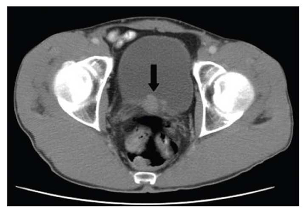
What is the irregular round filling defect in the right posterior aspect of the bladder?
The irregular round filling defect in the right posterior aspect of the bladder (arrow) represents a transitional cell carcinoma. There is thickening of the adjacent bladder wall, but not definite spread beyond the bladder.
How old is microscopic hematuria?
microscopic hematuria (<35 years old): can be performed according to bladder cancer suspicion
Which is better, CTU or cystoscopy?
Diagnostic performance of CTU was compared with cystoscopy in 177 patients. CTU performed better with 96.3% sensitivity, 86.4% specificity, 92.8% diagnostic accuracy, 92.9% PPV, and 92.7% NPV. The arterial acquisition phase diagnosed the lesions with the highest accuracy, and demonstrated 93.4% of all lesions [31].
Is CT more effective than MRI?
CT is faster and more cost-effective than MRI, but it is associated with the risk of ionizing radiation, high interobserver variability, and can neither differentiate bladder wall muscle layers, nor can it reliably distinguish T1 from T2 disease. Furthermore, its specificity and sensitivity are low for extravesical extension of early stage BCa and small metastatic lesions of BCa.[17, 21, 25, 26, 32–34].