Contents
Can a CT urogram detect bladder cancer?
· Can a CT scan miss bladder cancer? CT scans can provide important information about the urinary tract and bladder tumors. However, while some bladder tumors may be seen on a CT scan, others may not be apparent, such as smaller or flatter tumors. What to expect when having a CT scan. A CT scan is a painless procedure that is typically performed on an …
Can a CT scan always catch cancer?
· So, a doctor’s usage of a CT Scan to detect bladder cancer may show certain things. This may include the following: The formation of stones in the urinary tract can be seen. Infections Cysts Obstructions Tumors, and Traumatic Injuries However, a doctor can use a low or high-dose CT Scan to trace urinary stones.
Can an abdomen CAT scan diagnose bladder cancer?
A CT scan is a test that uses x-rays and a computer to create detailed pictures of the inside of your body. It takes pictures from different angles. The computer puts them together to make a 3 dimensional (3D) image. CT (or CAT) stands for computed (axial) tomography. You usually have a CT scan in the x-ray (radiology) department as an outpatient .
Does cancer always show up in a CT scan?
A CT scan of the kidney, ureters, and bladder is called a CT urogram. It can provide detailed information about the size, shape, and position of any tumors in the urinary tract, including the bladder. It can also help show enlarged lymph nodes that might contain cancer, as well as other organs in the abdomen (belly) and pelvis.
How accurate is CT scan for bladder cancer?
Computed tomography (CT) Multidetector (64-slice) CT scanning has provided the mainstay in radiological assessment. It has a reported sensitivity of 85% and specificity of 94% for the diagnosis of bladder cancers [11]. Detection is dependent on the morphology and size of the tumor.
Can a CT scan Miss bladder cancer?
Can a CT scan miss bladder cancer? CT scans can provide important information about the urinary tract and bladder tumors. However, while some bladder tumors may be seen on a CT scan, others may not be apparent, such as smaller or flatter tumors.
Do bladder tumors show up in CT scan?
Computed tomography (CT) scan It can provide detailed information about the size, shape, and position of any tumors in the urinary tract, including the bladder. It can also help show enlarged lymph nodes that might contain cancer, as well as other organs in the abdomen (belly) and pelvis.
What can a CT scan of the bladder show?
A CT scan combines x-rays with computer technology to create three-dimensional (3-D) images. These scans can show stones in the urinary tract, as well as obstructions, infections, cysts, tumors, and traumatic injuries. Imaging for urinary stone disease can be done with low or ultra-low dose CT scans.
What is the best test for bladder cancer?
Cystoscopy. Cystoscopy is the key diagnostic procedure for bladder cancer. It allows the doctor to see inside the body with a thin, lighted, flexible tube called a cystoscope. Flexible cystoscopy is performed in a doctor’s office and does not require anesthesia, which is medication that blocks the awareness of pain.
How do you rule out bladder cancer?
Tests for bladder cancer look for different substances and/or cancer cells in the urine. Urinalysis: One way to test for bladder cancer is to check for blood in the urine ( hematuria). This can be done during a urinalysis, which is a simple test to check for blood and other substances in a sample of urine.
Why would a urologist order a CT scan?
A CT urogram is used to examine the kidneys, ureters and bladder. It lets your doctor see the size and shape of these structures to determine if they’re working properly and to look for any signs of disease that may affect your urinary system.
Is cystoscopy necessary after CT scan?
While some bladder tumors may be found on a CT urogram or other imaging test, others will not. A urologist will often recommend a cystoscopy to evaluate the lower urinary tract (bladder/urethra) for a source of blood in the urine or to workup other urologic symptoms.
Why would a urologist order a cystoscopy?
During a cystoscopy, a urinary tract specialist (urologist) uses a scope to view the inside of the bladder and urethra. Doctors use cystoscopy to diagnose and treat urinary tract problems. These problems include bladder cancer, bladder control issues, enlarged prostates and urinary tract infections.
Is a CT urogram the same as a CT abdomen and pelvis?
A CT (computerised tomography) scan uses x-rays and a computer to create a detailed picture of the inside of the body. A scan of the urinary system may be called a CT urogram, CT IVP (intravenous pyelogram) or a triple-phase abdomen and pelvis CT – these are different names for the same test.
What organs are seen on a CT scan of abdomen and pelvis?
A CT scan of the abdomen and pelvis can help diagnose problems in the bladder, uterus, prostate, liver or bowels. This procedure is typically used to help diagnose the cause of abdominal or pelvic pain. It is also used to identify diseases of the internal organs such as: Appendicitis.
What is the difference between a CT scan and a CT urogram?
A CT urogram is a test that uses a CT scan and a special contrast medium or dye that a doctor injects into a vein. The contrast dye provides a high quality image to allow doctors to look at the urinary system and make a diagnosis.
Can bladder cancer symptoms come and go?
Symptoms often come and go, and are often not severe. The most common symptoms include the following: Hematuria (blood in the urine) — The most common sign of bladder cancer is blood in the urine (hematuria).
How accurate is a CT urogram?
CT urography was found to be as accurate as cystoscopy for patients with hematuria, (94.6% and 94.4% accurate, respectively). Both tests showed lower accuracy in the evaluation of patients with a history of urothelial cancer than in patients with hematuria, CT urography more so than cystoscopy (77.8% vs 84.8%).
Why do I need a cystoscopy after a CT scan?
While some bladder tumors may be found on a CT urogram or other imaging test, others will not. A urologist will often recommend a cystoscopy to evaluate the lower urinary tract (bladder/urethra) for a source of blood in the urine or to workup other urologic symptoms.
Can you have bladder cancer without blood in urine?
Among women with hematuria the rate of cancer was 1.7%, compared with 0.45% among those without hematuria. Among the 10 bladder cancer cases, six had no hematuria.
Medical History and Physical Exam
Your doctor will want to get your medical history to learn more about your symptoms. The doctor might also ask about possible risk factors, includi…
Transurethral Resection of Bladder Tumor (TURBT)
If an abnormal area (or areas) is seen during a cystoscopy, it will be biopsied to see if it is cancer. A biopsy is the removal of small samples of…
Biopsies to Look For Cancer Spread
If imaging tests suggest the cancer might have spread outside of the bladder, a biopsy might be needed to be sure.In some cases, biopsy samples of…

What does a CT scan of your pelvis tell you?
You might have a CT scan of your pelvis and tummy (abdomen). It can tell your doctor where the cancer is, how big it is and if the cancer has spread. Knowing this helps your specialist decide on the best treatment for you.
What is a CT scanner?
A CT scanning machine is large and shaped like a doughnut. You might have an injection of a type of dye called a contrast medium through a small tube (cannula) in your arm. You may: feel hot and flushed for a minute or two. have a metallic taste in your mouth.
What is a CT scan of the abdomen?
Knowing this helps your specialist decide on the best treatment for you. A CT scan is a test that uses x-rays and a computer to create detailed pictures of the inside of your body. It takes pictures from different angles.
What is a voiceover CT scan?
Voiceover: A CT scan helps your doctor make a diagnosis, decide about what treatment you need or find out if your treatment is working. This type of scan takes a series of x-rays and uses a computer to put them together. Before your scan you may need to drink either half a litre of water or a type of dye called a contrast medium.
What to drink before a scan?
Before your scan you may need to drink either half a litre of water or a type of dye called a contrast medium. This helps to make the scan clearer.
What is the number to call for cancer research?
For information and support, you can call the Cancer Research UK nurses on freephone 0808 800 4040. The lines are open from 9am to 5pm, Monday to Friday.
How to prepare for a CT scan?
You usually receive written instructions on how to prepare for your CT scan. Let the radiology department know if you’re taking a drug called Metformin. The dye (contrast medium or iodine) can interact with the metformin. Also, let them know if you have an allergy to the dye.
What is the best way to diagnose bladder cancer?
Cystoscopy. If bladder cancer is suspected, most doctors will recommend a cystoscopy. . A urologist uses a cystoscope, which is a long, thin, flexible tube with a light and a lens or a small video camera on the end. For details on how this procedure is done, see Cystoscopy.
What tests are used to check for bladder cancer?
These include the tests called NMP22 ® (or BladderChek ® ), BTA Stat ®, Immunocyt ® , and UroVysion ®, which are discussed in Can Bladder Cancer Be Found Early?

What is the blue light in a cystoscopy?
Fluorescence cystoscopy (also known as blue light cystoscopy) may be done along with routine cystoscopy. For this exam, a light-activated drug is put into the bladder during cystoscopy. It’s taken up by cancer cells. When the doctor then shines a blue light through the cystoscope, any cells containing the drug will glow (fluoresce). This can help the doctor see abnormal areas that might have been missed by the white light normally used.
What is the biopsy for bladder cancer?
A biopsy is when tiny pieces (called samples) of the abnormal-looking tissue are taken out and tested for cancer cells. If bladder cancer is suspected, a biopsy is needed to be sure of the diagnosis.
What is a physical exam for bladder cancer?
A physical exam can provide information about possible signs of bladder cancer and other health problems. The doctor might do a digital rectal exam (DRE), during which a gloved, lubricated finger is put into your rectum. If you are a woman, the doctor might do a pelvic exam as well.

How long does it take for a urine culture to show up?
It can take time for the bacteria to grow, so it may take a few days to get the results of this test.
How does ultrasound help with bladder cancer?
Ultrasound uses sound waves to create pictures of internal organs. It can be useful in determining the size of a bladder cancer and whether it has spread beyond the bladder to nearby organs or tissues. It can also be used to look at the kidneys. This is usually an easy test to have, and it uses no radiation.
Why do you need a bladder CT scan?
Some reasons to request a bladder CT scan can include incontinence, bladder masses, or signs of blockages. This test can also be requested if a doctor suspects the presence of stones in the bladder. It can be ordered if abnormalities are noticed on another imaging study like an ultrasound or X-ray, or if they are observed during an examination. Interpreting the images may require several days, depending on the staff at a facility.
How to get a picture of bladder?
Images of the bladder can be produced through use of a CT scan.
Can a bladder CT scan be done outpatient?
Imaging centers can usually offer a bladder CT scan as an outpatient procedure. The entire test, including checking in, acquiring the images, and being monitored for initial reactions if contrast was used, may take several hours. If a patient is already hospitalized for an existing medical problem, the test can be offered as an inpatient service, in which case people can transfer to the radiology department for the test and return to their beds when they are finished.
Is a bladder CT scan safe?
Risks associated with a bladder CT scan are low. Patients are exposed to some radiation, but it is kept as low as possible and the benefit of catching a problem outweighs the risk. Pregnant women may be advised to wait for testing if at all possible because of the increased risks for the developing fetuses. Some people experience allergic reactions to contrast agents, and it is important to discuss past allergy and medical history with a medical provider before starting the test. The technician may decide to use a different contrast agent or consider a bladder CT without contrast if this appears necessary.
What is the most common symptom of bladder cancer?
The most common (> 80%) clinical symptom of bladder cancer is gross hematuria, which confers a four times higher risk of malignancy compared with microscopic hematuria. Most patients with macroscopic hematuria, particularly those older than 50 years, should undergo both cystoscopy and CT urography [ 2 ]. Microscopic hematuria carries a much lower risk of malignancy and may be present in a sizeable percentage of persons without symptoms, making the decision to perform further investigation (CT urography or cystoscopy) largely dependent on a patient’s underlying risk factors [ 8 ]. Other than hematuria, potential patient symptoms include urinary frequency, urgency, repeated urinary tract infections, and urinary obstruction secondary to an advanced mass [ 2 ].
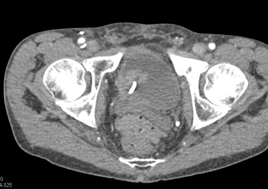
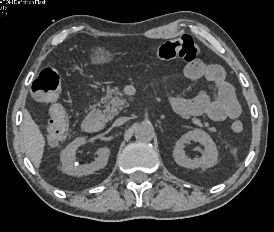
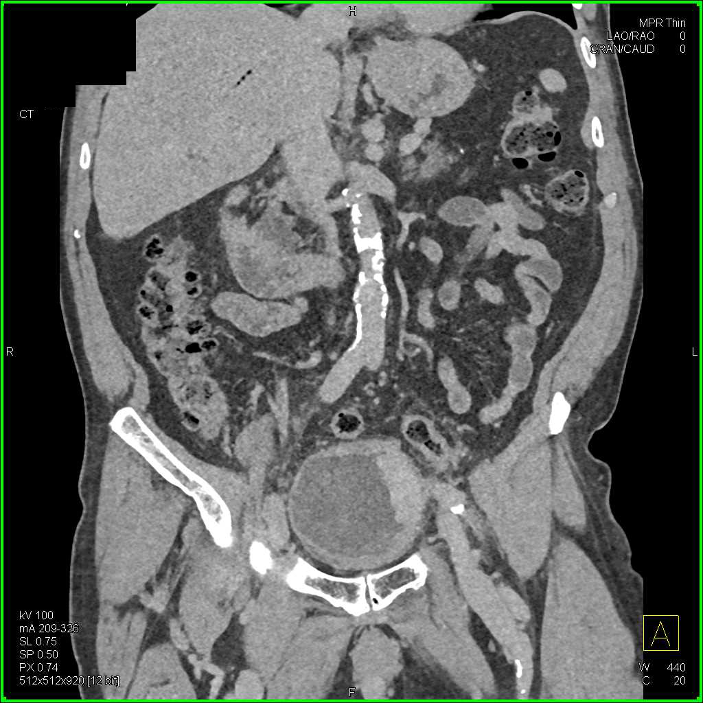
What is the arrow in a ct of the bladder wall?
A, Axial ( A) and coronal ( B) volume-rendered CT images show extensive polyplo id nodular thickening ( arrow, A) of bladder wall, in keeping with multifocal transitional cell carcinoma. Coronal image nicely shows extensive vascularity and enhancement associated with malignancy.
What is the CT image of a 30 year old man with multiple sclerosis?
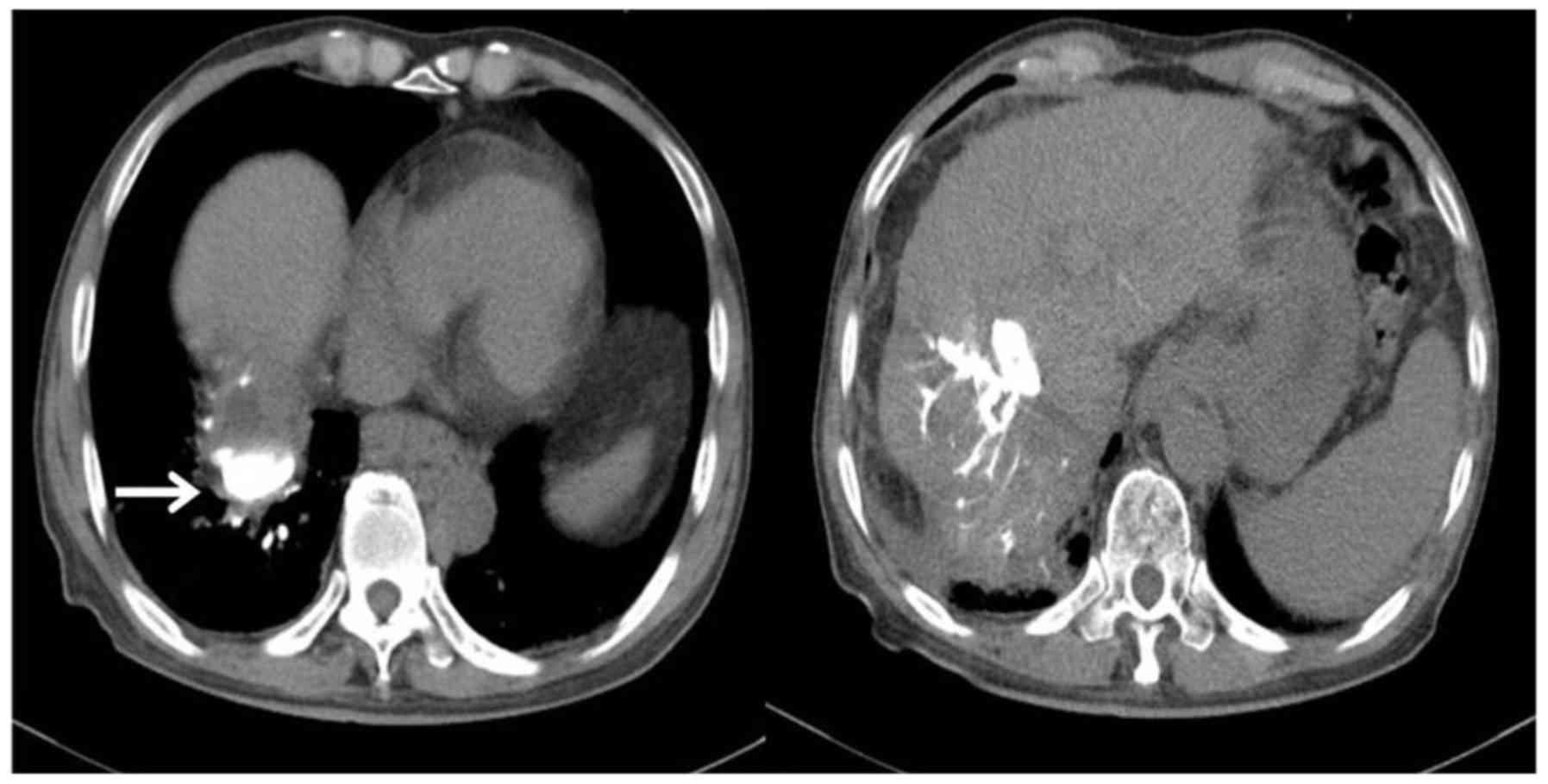
Fig. 6 —30-year-old man with history of multiple sclerosis. CT image shows multifocal bladder wall thickening and hyperenhancement. Given young age of patient and presence of multiple sclerosis and multifocal thickening of bladder, finding was originally thought to represent either cystitis or neurogenic bladder but was ultimately proved to represent diffuse infiltrating transitional cell carcinoma.
What is the role of CT urography?
The role of CT, CT urography in particular, has been well described in the literature with regard to the diagnosis of upper urinary tract tumors (i.e., tumors of the ureters and pelvicalyceal system) [ 1 ]. The role of CT in the imaging of patients with hematuria has become well established, and CT urography is regularly used in both the out-patient and emergency settings.
What are the techniques used in urography?
In the performance of CT urography, a number of ancillary techniques have been described, including oral hydration, IV hydration, IV administration of diuretics, abdominal compression, and prone positioning [ 11, 13 – 16 ]. For the most part, these techniques have been described in the literature with regard to improving the efficacy of upper uri-nary tract distention, and their effect on the detection of bladder malignancies has not been specifically discussed. Although some of these techniques are unlikely to have a substantial effect on bladder distention, three of these techniques are likely to improve detection of a bladder malignancy: oral hydration, IV hydration, and IV administration of diuretics. These techniques can improve excretion into and distention of the collecting system and ultimately improve bladder distention. Moreover, all three result in dilution of the excreted contrast material, potentially improving the ability to identify subtle lesions.
Is cystoscopy more sensitive than urography?
Although there is little doubt that cystoscopy is the most sensitive, specific, and reproducible method for identification of bladder malignancies, the diagnostic performance of CT is better than is commonly thought: In a study by Turney et al. [ 8 ], 200 patients with hematuria underwent both cystoscopy and CT urography. Bladder malignancy was found in 24% of the patients, and the sensitivity and specificity of CT urography were 0.93 and 0.99 (positive predictive value, 0.98; negative predictive value, 0.97). Similarly, Sadow et al. [ 17] conducted a study that included 838 patients who had undergone CT urography and cystoscopy within 6 months of each another and found that CT urography had a sensitivity and specificity of 79% and 94% (positive predictive value, 75%; negative predictive value, 95%). In another study, Kim et al. [ 18] found CT urography effective in the diagnosis of recurrent tumors in patients who had previously undergone transurethral resection of a bladder tumor. It is undoubtedly true that the diagnostic performance of CT will vary dramatically depending on the technique used and degree of bladder distention, thus explaining the variable diagnostic performance from one study to another. A dedicated CT urography technique with multiphase protocol will clearly be more sensitive for a subtle tumor than a routine screening examination performed in the emergency department for suspected abdominal pain. Nevertheless, the results of these studies suggest that if technique is optimized, subtle malignancies can be identified reasonably accurately at imaging.

Does a split bolus reduce bladder distention?
Given that a smaller amount of IV contrast material is ultimately excreted in the latter two protocol options, it is not surprising that use of the split-bolus and triple-bolus techniques ultimately reduces the degree of uri-nary bladder distention on delayed phase images and may limit identification of a subtle filling defect. However and most important, both the split-bolus and the triple-bolus techniques were designed primarily to reduce radiation dose by combining the different phases of acquisition. As a result, each of these two protocols may markedly limit sensitivity for bladder malignancies if early phase images with unopacified urine distending the bladder are not acquired [ 1, 10 – 12 ].
What is a CT scan?
A CT scan is a fast, painless, and non-invasive medical imaging test used to screen for cancer.
Why do we need CT scans?
One example of a situation that may need regular CT scans is undergoing cancer treatment . If the cancer is in an area such as the lungs, regular CT scans can help practitioners see how the treatment is working. In this case, the patient’s cumulative radiation dose increases.
How long does it take to get a CT scan?
Depending upon the part of the body being scanned, it may take anywhere from 10 to 30 minutes. During the scan, you may be asked to hold your breath for a few seconds.
Which is better, MRI or CT?
In some cases, an MRI is much better at showing certain cancers than a CT scan.
How does a round scanner work?
Inside the round scanner, an X-ray tube rotates and emits a thin beam of X-rays at different angles that quickly pass through the patient’s body and are received at the opposite end. The signals are then passed to a computer where special software creates detailed images of the inside of the body.
Does a CT scan have ionizing radiation?
A CT scan uses ionizing radiation that may increase the risk of developing cancer. An MRI scan does not have harmful ionizing radiation.
Is a single imaging test 100% accurate?
No single imaging test is 100% accurate in detecting abnormalities. There may be a misdiagnosis due to the quality of the scan or due to the expert reading the scan.